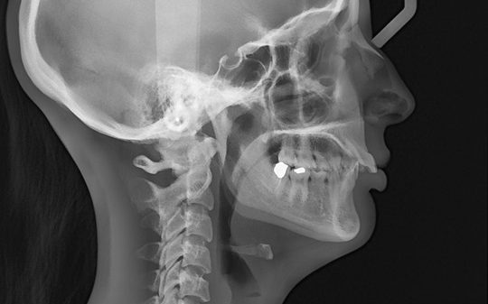
The cephalometric X-ray is a unique tool, which enables the dentist to capture a complete radiographic image of the side of the face. X-rays in general offer the dentist a way to view the teeth, jawbone and soft tissues beyond what can be seen with the naked eye. Cephalometric X-rays are extraoral, meaning that no plates or film are inserted inside the mouth. Cephalometric and panoramic X-rays display the nasal and sinus passages, which are missed by intraoral bitewing X-rays.
Cephalometric X-rays are usually taken with a panoramic X-ray machine. The adapted machine will have a special cephalometric film holder mounted on a mechanical arm. An X-ray film is exposed to ionizing radiation in order to provide the dentist with pictures of the entire oral structure.
Cephalometric X-rays are not as common as “full sets” or bitewing X-rays, but they serve several important functions:
- Provide views of the side profile of the face.
- Provide views of the jaw in relation to the cheekbone.
- Provide information about “bad bites” or malocclusions.
- Allow measurement of the teeth.
- Identify fractures and other injuries to the teeth and jawbone.
- Assists in orthodontic planning.
How are cephalometric X-rays taken?
Cephalometric X-rays are completely painless. The head is held with plastic ear rods to stabilize and align the head at a specific distance from the x-ray source and film.
After capturing cephalometric X-rays, the dentist will be able to see a complete side profile of the head. This can assist in orthodontic planning, and allow an immediate evaluation of how braces might impact the facial profile and teeth.